
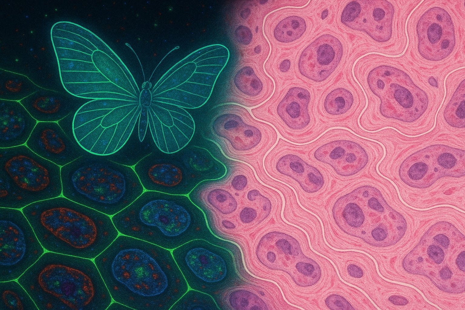
The Immunity Butterfly Effect
How spatial biology and causal AI are redefining drug discovery

TL;DR
1. Why Now?
We finally have the tools to decode human biology at unprecedented depth and breadth. Nowhere is this more urgent (or more complex) than in immunology, where small changes in dynamic, mobile immune cells can trigger a butterfly effect, leading to major consequences for patient outcomes. New computational approaches are emerging to make sense of the chaos.
2. The Rise of Perturbation Modeling
Imagine asking “what if?” without lifting a pipette. Perturbation models simulate how cells would respond to gene edits, drugs, or environmental shifts, guiding experiments and surfacing therapeutic hypotheses. When paired with spatial context, this modeling gets closer to cause, not just correlation.
3. The Promise and Pitfalls of Spatial Transcriptomics
Spatial transcriptomics has drawn major hype and investment. Its biggest promise (“big data” at scale without losing spatial context) has materialized with growing panel sizes, peaking with full-transcriptome. But behind the scenes, users face noisy data, inconclusive results, and high assay and instrumentation costs.
4. Spatial Proteomics: A Clearer Window
By contrast, high-plex protein imaging offers a direct, high-fidelity readout of cell status, including its identity and function. These methods detect key functional markers at subcellular resolution, capturing rare immune niches and actionable patterns that transcriptomics often misses.
5. Case Study: Noetik — Simulating Tumor–Immune Interactions
Noetik is building a spatial foundation model for NSCLC, trained on vast, multimodal data. Its transformer model (OCTO) can run virtual perturbation experiments inside tumor sections, predicting how immune cells might respond to environmental or genetic changes. It’s scale-first, simulation-centric biology.
6. Case Study: Voyant Bio — Mapping Immune Cell Interactions
Voyant maps conserved immune resistance patterns across cancers using high-plex imaging and immune cell interaction networks. It validates model predictions in ex vivo patient tumor fragments, then engineers biologics that act on the most promising cell interactions. From insight to drug in one loop.
7. Two Paths, One Goal
Noetik bets on simulation scale and model generalization; Voyant bets on biological causality and fast wet-lab feedback. Both aim to crack immune resistance. Their differing strategies reflect complementary views of how to turn complex spatial biology into curative immunotherapies.
8. Strategic Outlook
The future lies at the intersection of spatial omics, causal AI, and protein-level precision. Spatial transcriptomics may be the flashier field, but spatial proteomics is closer to clinical utility. I argue that platforms that pair direct deep protein measurement with fast functional validation are best positioned to deliver meaningful ROI, and real cures.
Why Now?
Right now, we have an incredible set of tools to finally tackle the complexity of human biology in ways that could lead to real cures. But which tools are most useful, and how do we apply them?
One of the most exciting and rapidly growing areas of investigation is the immune system’s contribution to many, if not most, physiological changes (e.g., aging) and pathological conditions. Single-cell methods, spatial omics, and AI have reshaped our understanding of the immune system in just a few years. Studying the immune system is especially challenging because it’s not static. Immune cells are highly diverse, can exist in many different states, and are constantly moving and interacting in complex ways. This makes it a much harder target than something like cells from any organ tissue, which are more homogeneous and stay in place.
What ties all of this together is the butterfly effect: small changes in biology can lead to big differences in whether a disease gets worse or a therapy works. Figuring out how these systems behave and respond is one of the biggest challenges in biology today.
In this piece, I’ll walk through the tools that are helping us map and make sense of this complexity, and spotlight some of the companies leading the way.
The Rise of Perturbation Modeling
One exciting new approach in biology is perturbation modeling, a way to predict how cells might react to different changes, like gene edits or drug treatments, without running actual experiments. And when you combine it with spatial tools that keep the tissue’s layout intact, it becomes even more powerful.
Take Perturb-map, for example. It used about 50 CRISPR guides targeting immune-related genes in a mouse lung cancer model. By layering protein imaging, researchers could see how knocking out each gene changed where CD8 T cells ended up in the tumor.
Another tool, Celcomen, modeled what would happen if you knocked out EGFR in lung cancer tissue. It predicted shifts in macrophage and T cell patterns, and those predictions held up when tested with advanced imaging.
Ultimately, these models help guide experiments, generate hypotheses, and forecast transcriptomic shifts, even in unmeasured cell states or conditions.
The Promise and Pitfalls of Spatial Transcriptomics
Supporting this are spatial omics techniques like Slide-seq, seqFISH, MERFISH, and DBiT-seq. These methods resolve molecular profiles directly in tissue, capturing how local environments and cell–cell relationships influence cellular responses. Sequencing-based tools like Slide-seq offer broad transcriptome coverage at ~10 μm resolution, while imaging-based platforms like seqFISH and MERFISH provide subcellular detail with more limited gene panels. This spatial dimension is key to understanding how perturbations ripple through tissue architecture.
Spatial transcriptomics (ST) has generated enormous excitement, with projections placing the market near $1 billion by 2030. But while the technology holds real promise, real-world adoption has run into significant technical and practical limits.
Core platforms like 10x Visium, NanoString GeoMx, CosMX, and Slide-seq are costly (~8-12K/slide), complex, and come with trade-offs between resolution, sensitivity, and completeness.
Add to that common issues like dropouts, ambient RNA contamination, and blurry cell boundaries, and the result is extremely noisy data. These challenges make it difficult to distinguish important cell states, or to accurately map cell–cell interactions.
Also, RNA is only an indirect proxy for cell state. Many important proteins are regulated post-transcriptionally. T cells, for example, often show poor correlation between mRNA levels and actual protein expression. Key immune markers (CD4, CD8, PD-1, PD-L1) are typically measured by protein assays like IHC or IF, not RNA. Studies have shown that PD-L1 mRNA levels often don’t reflect its protein abundance.
Another major limitation: you often can’t get both high-quality spatial RNA and protein data from the same tissue section. Many ST workflows (e.g., 10x Visium) require fresh-frozen or FFPE sections on specialized slides, and their processing steps can destroy protein epitopes. This forces researchers to perform protein imaging on adjacent sections, losing precise spatial alignment. Some emerging multi-omics approaches try to address this, but they’re even more expensive and often low-plex.
In short, while spatial transcriptomics is a powerful tool for exploratory research and hypothesis generation, it remains noisy, indirect, and expensive. It excels at mapping broad gene-expression landscapes, but often falls short when used alone for precise cell phenotyping or interaction discovery.
Spatial Proteomics: A Clearer Window
In contrast to spatial transcriptomics, multiplexed protein imaging (spatial proteomics) offers a more direct and high-resolution view of cell identity and function. Platforms like cyclic immunofluorescence (CyCIF, IBEX, PhenoCycler, Comet, InSituPlex), mass spectrometry–based imaging (IMC, MIBI), and multispectral imaging (Opal, Orion) now routinely achieve true single-cell and subcellular resolution on standard tissue sections.
For example, an early published multiplex study demonstrated 60 protein markers detection on a single FFPE slide using sequential fluorescent staining. This approach is now challenged by the need to expand the size of the panels (up to 100 markers) and their depth (including niche-specific and unique markers). Automated multiplex IF protocols now commonly detect 6 to 20+ proteins per section, offering highly reproducible, high-fidelity cell profiling. Crucially, in these larger panels, each marker corresponds to a well-validated antibody, yielding direct measurements of functional markers (rather than just cell type markers) with minimal ambiguity. Because these are protein-level readouts, they capture cell states and spatial patterns more reliably than mRNA alone.
Beyond static lineage or checkpoint markers, truly dynamic protein events can report when and where immune cells are actively engaging their targets. A classic example is NFAT: upon TCR triggering by an antigen–MHC complex on a dendritic cell, NFAT rapidly translocates from the cytoplasm into the nucleus. By imaging NFAT localization, you get a direct, single-cell readout of early TCR signaling and antigen engagement in situ.
To achieve even finer resolution, proximity ligation assays (PLA) allow detection of direct receptor–ligand interactions. In PLA, pairs of antibodies - against proteins such as TCR and peptide-MHC or PD-1 and PD-L1 - carry DNA “tags” that only ligate and amplify when the two proteins come into molecular contact. Coupled with multiplex IF, PLA reveals exactly which cells form synapsing, how frequently these interactions occur, and in which micro-environmental niches, information impossible to infer from gene expression or single-color stains alone.
Spatial proteomics is also more scalable and cost-effective. A well-optimized panel of 20 markers can be run on multiple slides per week using automated stainers. Costs per datapoint are lower, implementation is faster, and workflows align with existing diagnostic practices. Sequencing is unnecessary, and image output can quickly undergo QC to identify and account for artifacts. Companies like Akoya (PhenoCycler), Ultivue, NanoString (PhenoMosaic), and PerkinElmer (Opal) are commercializing these workflows, helping to drive rapid growth in the spatial proteomics market.
This rapidly growing field bridges diverse areas of expertise that previously had few points of intersection, enabling collaboration among physicians, pathologists, image analysts, ML/AI algorithm developers, basic science researchers, technology innovators, and commercial platform providers. These collaborations drive rapid and extraordinary developments with a multifaceted approach, advancing our understanding of human health and disease.
Next, we’ll dive into two case studies of companies in the spatial biology space.
Case Study: Noetik — Simulating Tumor–Immune Interactions
Noetik is building a next-generation foundation model for spatial biology, with an initial focus on non-small cell lung cancer (NSCLC). Their core belief is that richly paired, high-dimensional data, fed into a self-supervised model, can eventually simulate complex tissue biology in silico. Rather than running thousands of wet-lab perturbation experiments, researchers could ask counterfactual questions directly within a virtual tumor microenvironment.
At the center of Noetik’s strategy is a massive data-generation effort: over 1,000 NSCLC tumors are being profiled across tissue microarray (TMA) cores. Each core is measured in four tightly integrated ways, protein-level phenotyping via 16-plex immunofluorescence (mIF), single-cell spatial transcriptomics using 1,000-plex CosMx, H&E histology for morphology, and DNA mutation panels for genomic context.
The computational centerpiece is OCTO, a multimodal transformer trained via self-supervised learning. OCTO learns to reconstruct missing data channels, such as predicting the full 16-marker mIF image from a partially masked input or inferring gene expression from morphology. OCTO’s most powerful capability lies in counterfactual simulation. Researchers can place a “virtual cell” anywhere in a tissue section and simulate what happens when its environment is perturbed. For example, OCTO can generate TLS (tertiary lymphoid structure) heatmaps that highlight known targets like LTB/LTBR or CCL19, suggest neighborhood swaps to study cell–cell influence (e.g., replacing a B cell’s neighbors with CD4 T cells), or run virtual target screens, dropping a CD8 T cell into any niche, silencing tumor genes, and ranking the changes in cytotoxic signatures like NKG7.
Still, the platform faces clear limitations. The current 16-plex mIF panel puts a ceiling on immune resolution, any phenotype not marked by those proteins may be effectively invisible unless spatial transcriptomics is also applied. But that approach is costly, not scalable to all samples, and typically can’t be run on the same slide, meaning the system may be analyzing different cells. OCTO’s predictive power is also closely tied to its training context: because it was trained on NSCLC data, applying it to other tissue types could reduce accuracy unless retrained with new examples, requiring access to large-scale cohorts of precious patient tissue. Also, rare but biologically critical cell states may be poorly represented, potentially missing butterfly effect dynamics that drive key tumor–immune interactions. Finally, the predictions are being tested in a mouse model, a step that has held back many immuno-oncology programs because results often don’t carry over well to real patients.
Looking ahead, Noetik’s in-silico perturbation engine shows real promise for hypothesis generation, immune interaction modeling, and spatial target discovery in lung cancer. But expanding beyond NSCLC will require additional multimodal datasets, and careful model adaptation to new tumor contexts. If successful, Noetik’s approach could transform how researchers simulate tissue biology, making spatial prediction, not experimentation, the new starting point for biological insight.
Case Study: Voyant Bio — Mapping Immune Cell Interactions
Voyant Bio is taking a bold approach to immuno-oncology. Rather than stopping at spatial atlases or descriptive maps, we’re focused on decoding and actively reprogramming resistance to immunotherapy across multiple solid tumor types. Our central thesis is that spatial discoveries must tie directly to function and therapeutic design, turning insight into action. To that end, Voyant is building a full-stack platform that integrates high-resolution imaging, self-supervised graph-based cell-cell interaction modeling, and patient-derived functional assays to drive first-in-class biologic therapies.
At the heart of Voyant’s strategy is high-plex cyclic immunofluorescence (cycIF), which pushes beyond conventional limits to measure 50+ protein markers per tissue section. This allows us to resolve nuanced and previously inaccessible cell states. We apply this platform across multiple cancer types to identify conserved mechanisms.
Voyant transforms this rich spatial data into cellular interaction graphs. These graphs are then analyzed using proprietary machine learning approaches, which allow the system to learn not just which cells are where, but how they communicate, and which interactions might drive therapeutic response or failure.
One of the most striking findings from such approach is the discovery of a conserved “immune triad,” a coordinated presence of progenitor CD8 T cells, activated dendritic cells, and CD4 T helper cells, that reliably explains response to PD-1 checkpoint blockade across tumor types. Unlike traditional markers such as CD8 infiltration, this triad captures upstream signaling cues that govern T cell fate long before exhaustion sets in. This rare constellation truly represents a butterfly effect, where subtle, early immune coordination triggers a cascade that ultimately determines therapeutic success or failure. These insights open new windows for intervention, aimed at preserving or restoring early immune activity.
Crucially, Voyant doesn’t stop at modeling. It validates its predictions using patient-derived tumor fragments (PDTFs), live micro-tumors from patients that can be perturbed ex vivo to measure the functional response of the exact cell states flagged by the model. This is a rare human-relevant readout, and the FDA has formally recognized such systems to be suitable for drug development. These insights are already feeding into drug discovery - Voyant is already engineering novel multi-specific therapeutics targeting key interaction patterns.
Voyant’s advantages are clear: its high-plex imaging uncovers rare but critical immune states that are routinely missed by lower-plex protein methods and even higher-plex transcriptomics. Our ML/AI framework captures true intercellular communication (not just spatial proximity) enabling a deeper understanding of tissue-level immune logic. And our closed-loop, active-learning system (spanning discovery, perturbation, and therapeutic development) dramatically accelerates the traditional hypothesis-to-therapy cycle.
We’ve already leveraged such approach to uncover a novel interaction (the triad) and are actively capitalizing on this concrete finding. Our team has designed, manufactured, and advanced the first therapeutic candidates targeting this interaction into pre-clinical testing, putting us ahead of the curve.
Of course, challenges remain. Cyclic IF is slower and more costly than standard mIF, but we argue that the increased resolution and biological richness more than justify the investment. The model’s accuracy also depends on diverse, well-annotated training sets, a challenge Voyant is tackling through multi-institutional partnerships and by engaging physician-scientists to ensure quality metadata.
In the long run, Voyant is positioning itself not as a tools or atlas company, but as a drug developer powered by deep spatial insight. If its biologics prove effective in the clinic, it could mark a major turning point in how spatial biology fuels precision immunotherapy.
Two Paths, One Goal
Noetik and Voyant Bio both lead the spatial biology field by leveraging high-plex spatial imaging and advanced AI architectures to decode the intricate tumor-immune or immune-immune cross-talk. Yet, they take fundamentally different routes to solving a similar problem: converting immunotherapy non-responders into durable responders.
Noetik is building a foundation model centered on NSCLC, applying multimodal transformers to simulate the tumor microenvironment and perform in-silico perturbation experiments at scale. Their strength lies in the pairing of large, richly annotated datasets (protein, RNA, histology, DNA) with self-supervised learning to generate predictive maps of cellular response.
Voyant, by contrast, focuses on graph-based causality across multiple tumor types. Their models learn from rare but critical cell-cell interactions, captured through high-plex cyclic IF, and then validate mechanisms directly in patient-derived micro-tumors. These functional readouts immediately inform the design of novel multi-specific biologics, creating a fast feedback loop from spatial insight to drug candidate.
While Noetik represents the simulation-at-scale bet, Voyant prioritizes biological causality and wet-lab validation. Their complementary strengths, Noetik’s massive, unified model and Voyant’s precise, real-world perturbation loop, underscore two viable approaches to next-gen immunotherapy.
Strategic Outlook
The convergence of spatial omics and perturbation modeling, driven by causal inference, generative AI, and graph-based learning, is turning spatial biology from a descriptive tool into a predictive engine for drug discovery.
While spatial transcriptomics has captured significant attention, current implementations often fall short in practical settings. ST platforms tend to be costly, low-resolution, and indirect, with noisy outputs that require heavy denoising and often underperform in phenotyping or predictive tasks. Their R&D application remains largely exploratory, with many systems still confined to academic use.
By contrast, multiplexed spatial proteomics, especially imaging-based, rests on a more established foundation. It directly measures functional protein markers, aligns with clinical practices like IHC/IF, and scales more cost-effectively per datapoint. It allows researchers to directly count and locate functional cell states and their interactions. These data not only reveal who is present, but who’s talking to whom, and with what consequence. Already, high-plex spatial protein data are influencing real clinical decisions, identifying immune niches and targets, predicting checkpoint response, and informing patient stratification.
Future developments in the field likely include:
High-throughput ex vivo screening and closed-loop biologic engineering
Foundation models that generalize across tissues, diseases, and species
Multi-modal perturbations spanning proteomic and epigenomic axes
Multiple, spatially resolved-omics on a single slide
Bottom Line
High-plex, protein-level spatial data meets causal AI and fast experimental validation is the combination most likely to translate complex tumor immunology into predictive biomarkers, targeted therapies, and ultimately, durable cures.